Education Chart of Biology for Human Cell Diagram Best Acupuncture llc
A cell is the smallest living thing in the human organism, and all living structures in the human body are made of cells. There are hundreds of different types of cells in the human body, which vary in shape (e.g. round, flat, long and thin, short and thick) and size (e.g. small granule cells of the cerebellum in the brain (4 micrometers), up to the huge oocytes (eggs) produced in the female.
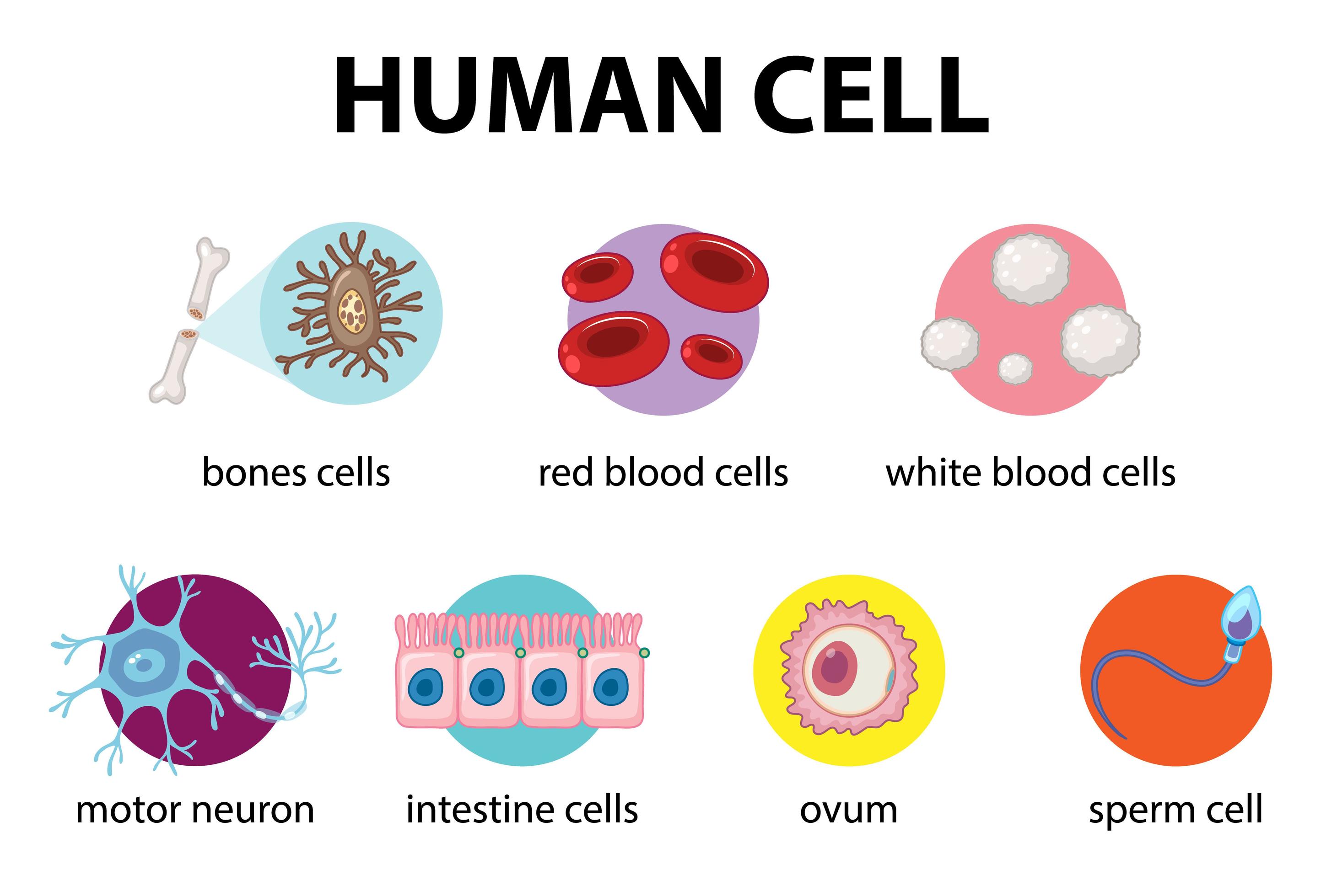
Diagram of human cell for education 1762228 Vector Art at Vecteezy
As human skin needs water and food to keep the cells in shape, the plant cells need sunlight to make their energy and sugar. Human cells do not make energy, plant cells do. In fact, humans are animals that eat energy and plants are a good food because they have energy stored. Also, plants don't need more than the sun and water to get energy.

Update 57+ human cell drawing latest nhadathoangha.vn
Human Cell - Properties, Diagram, Parts, Pictures, Structure. By Thulasi Ram. The cell is the basic unit of any living organ and it is the organ that replicate on its own determining growth. The cell does not need any other triggering element for its multiplication since it is self contained. Cell was first discovered by Robert Hooke in the.
human cell types Anatomy System Human Body Anatomy diagram and
Cell Structure. Ideas about cell structure have changed considerably over the years. Early biologists saw cells as simple membranous sacs containing fluid and a few floating particles. Today's biologists know that cells are infinitely more complex than this. There are many different types, sizes, and shapes of cells in the body.

Human Cell Diagram Cell diagram, Human cell diagram, Plant and animal
?µ 'šÔ "0nâc çûÏL? .§×Hg$Í \EQK‹)yI¢ ÛqmñÔOü] '°A€ Jb ûß›Zï†ÿ{Y[¹|$eÊÖRÎ$±óçÞûÞêu7Zj€d rJ w¶ŠàŸ ÇTqÖÜ÷Ú Ñ.

Pin by james paterson on A (growing) list of people, places and things
Interactive guide to stem cells and cell biology with 3D models and real microscopy data of GFP labeled hiPSCs.

Explain the nucleus of a cell with a neat labeled diagram Science
Detailed diagram of lipid bilayer of cell membrane. The cell membrane, or plasma membrane, is a selectively permeable biological membrane that surrounds the cytoplasm of a cell.. Human cancer cells, specifically HeLa cells, with DNA stained blue.
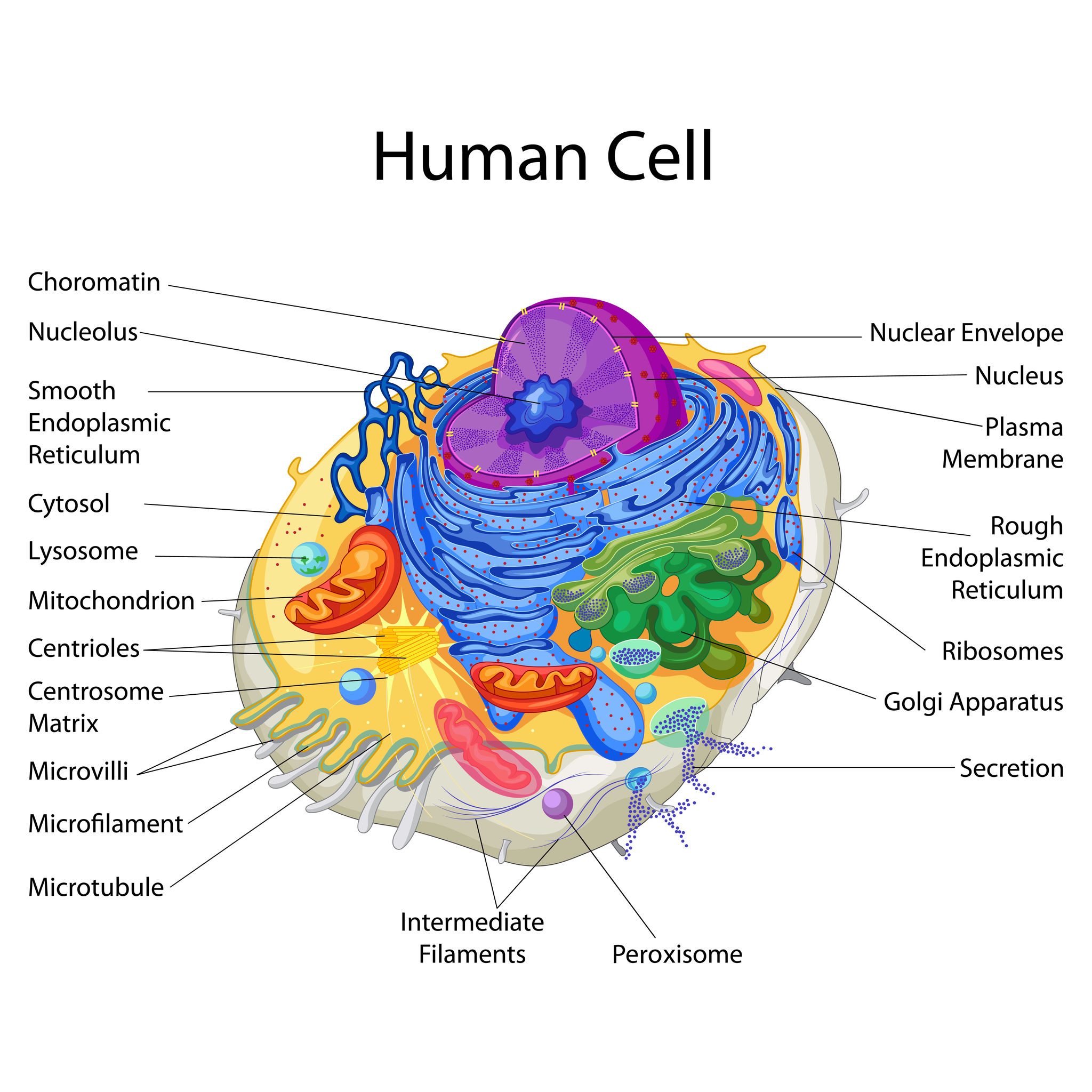
Human Cell Diagram 6406474 Vector Art at Vecteezy
To learn more about cells, check out our free Human Cell eBook! Cells can be divided into four groups: somatic, gamete, germ, and stem. Somatic cells are all the cells in the body that aren't sex cells, like blood cells, neurons, and osteocytes. Gametes are sex cells that join together during sexual reproduction. Germ cells produce gametes.
Cells Haleo
Cells. The most basic parts of the human machine are cells—an amazing 100 trillion of them by the time the average person reaches adulthood! Cells are the basic units of structure and function in the human body, as they are in all living things. Each cell carries out basic life processes that allow the body to survive.

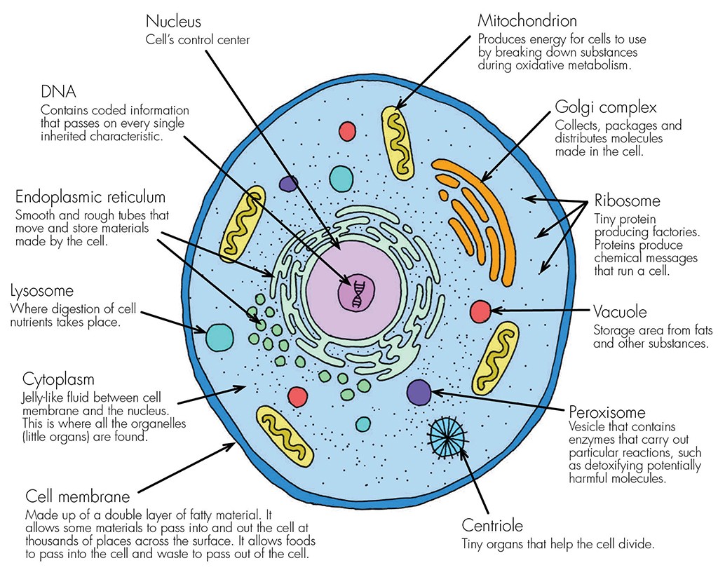
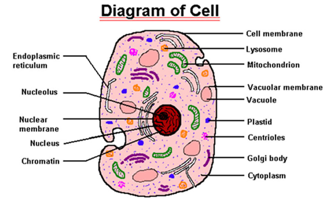
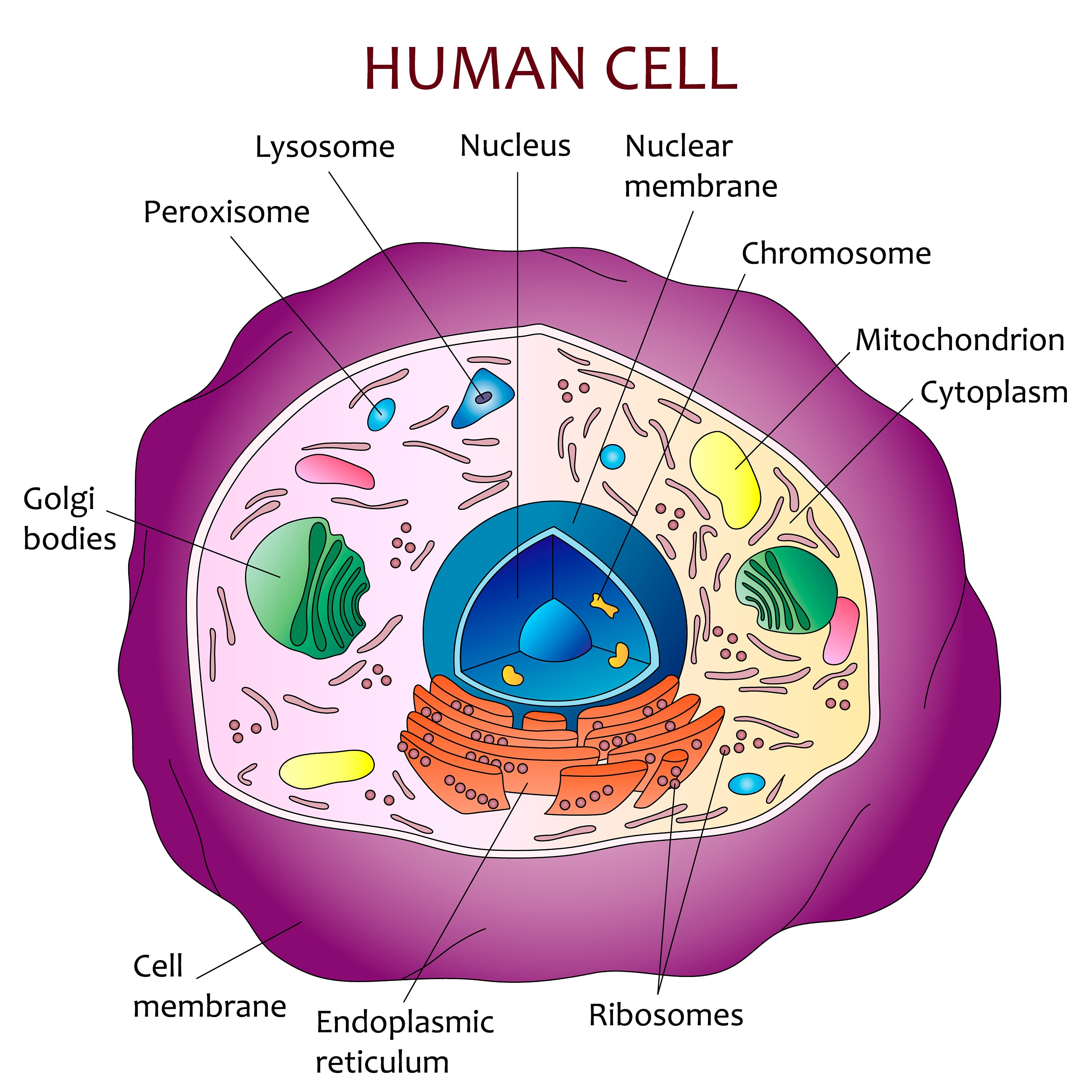
Human Cell Diagram, Parts, Pictures, Structure and Functions
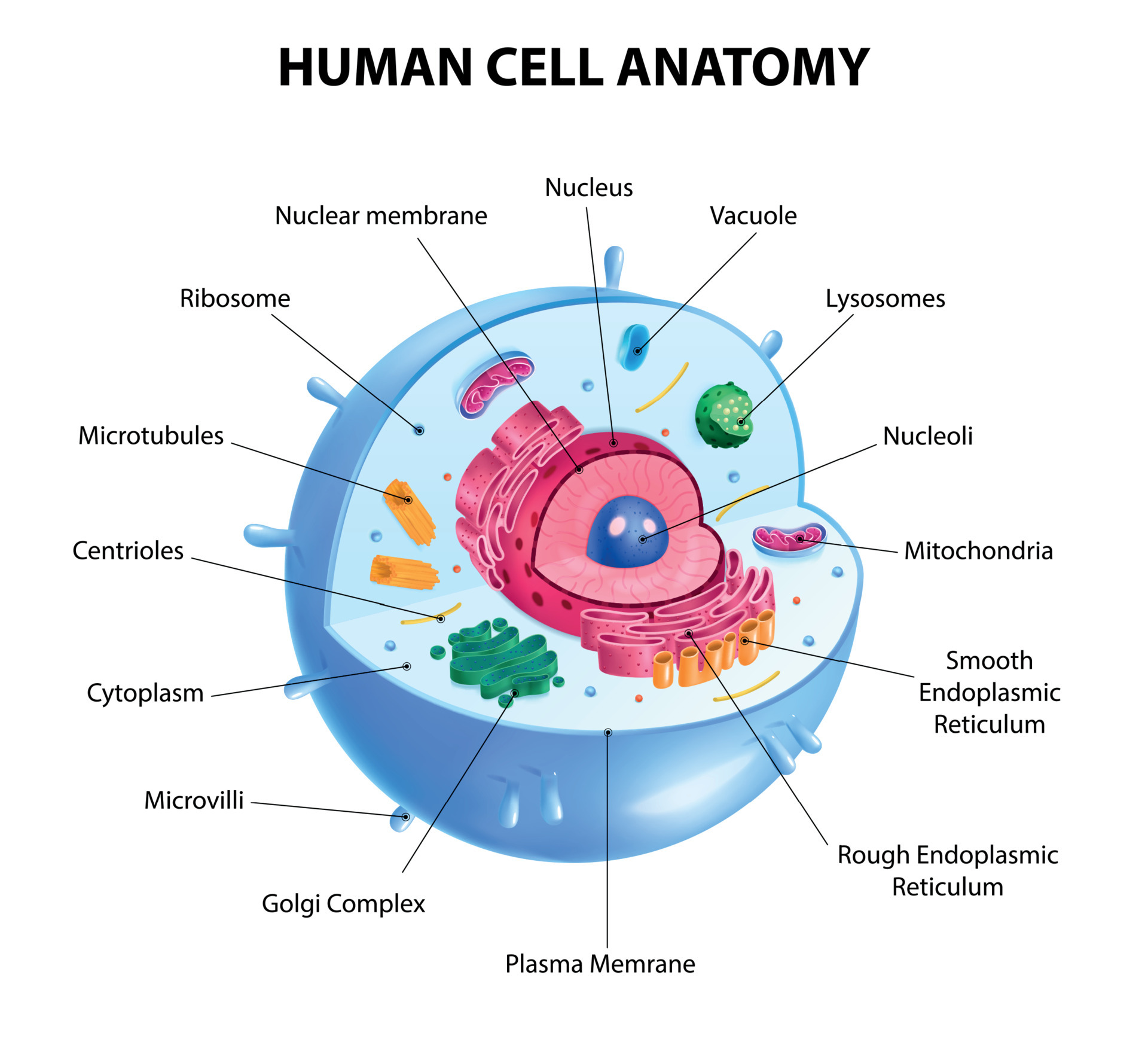
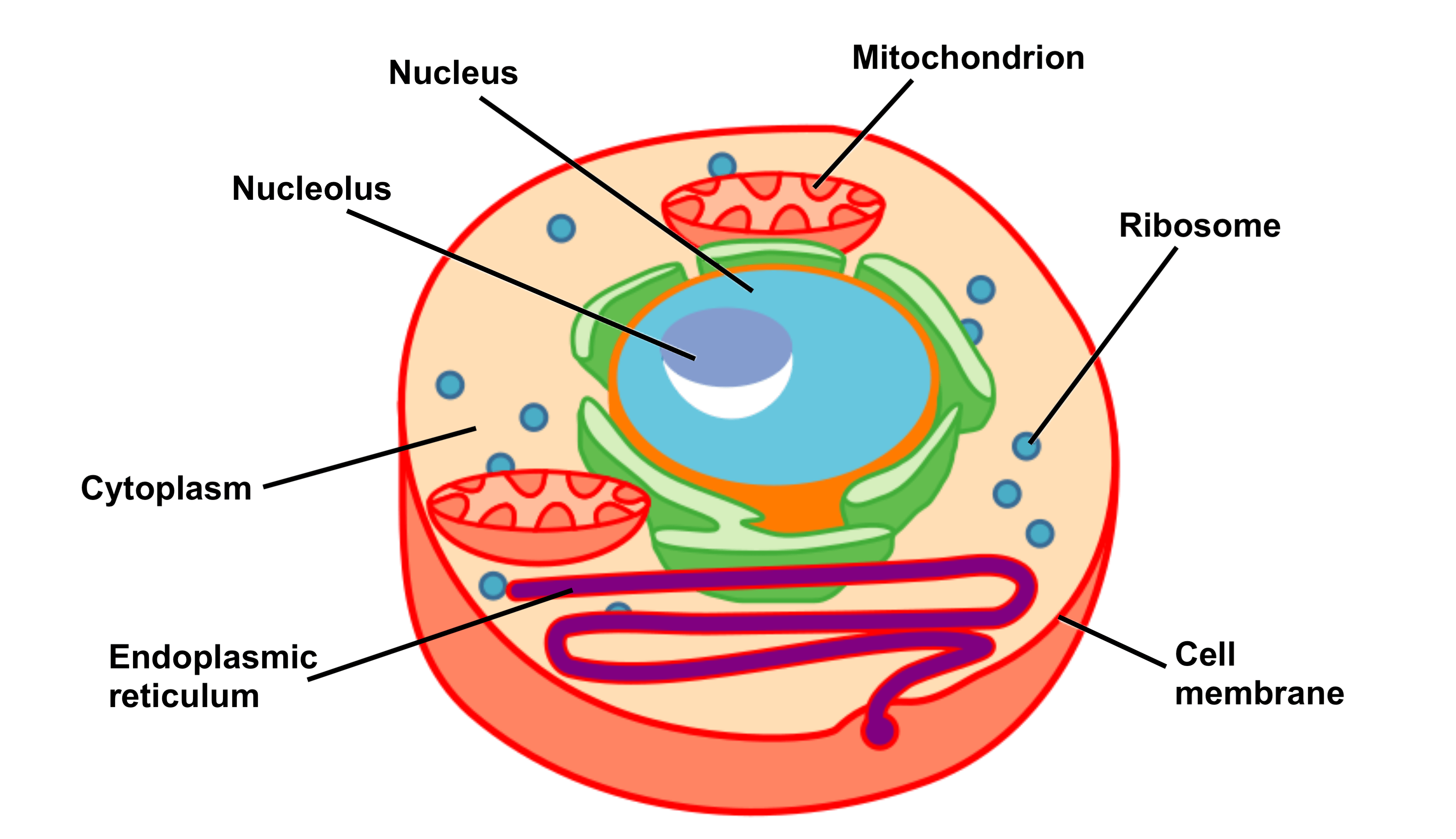
Diagram 2: The human cell membrane. Image Source: www.curezone.org. Cytoplasm/Protoplasm. It is the fluid inside the cells, which allow a number of cell organs to float inside the cell. It contains a nucleus surrounded by a nuclear membrane. It consists of molecules, enzymes, fatty acids, sugar, and amino acids. (3)
Education 645 High School Biology
The Human Cell Atlas is likely to impact almost every aspect of biology and medicine, leading to a richer understanding of life's most fundamental units and principles.Some examples of what a cell atlas is helping scientists and physicians do: Provide a reference map for comparing related cells and identifying new cell types, states (behaviors), and how cells transition among them.
Human cell diagram Etsy
How many cells are in the human body.. Now you see it in this diagram right over here. This is not a common feature to all cells but the only reason why I'm mentioning it in this video is officially, the cytoplasm does not include the stuff inside the nucleus. In a eukaryotic cell, that is called the nucleoplasm but we'll talk more about.
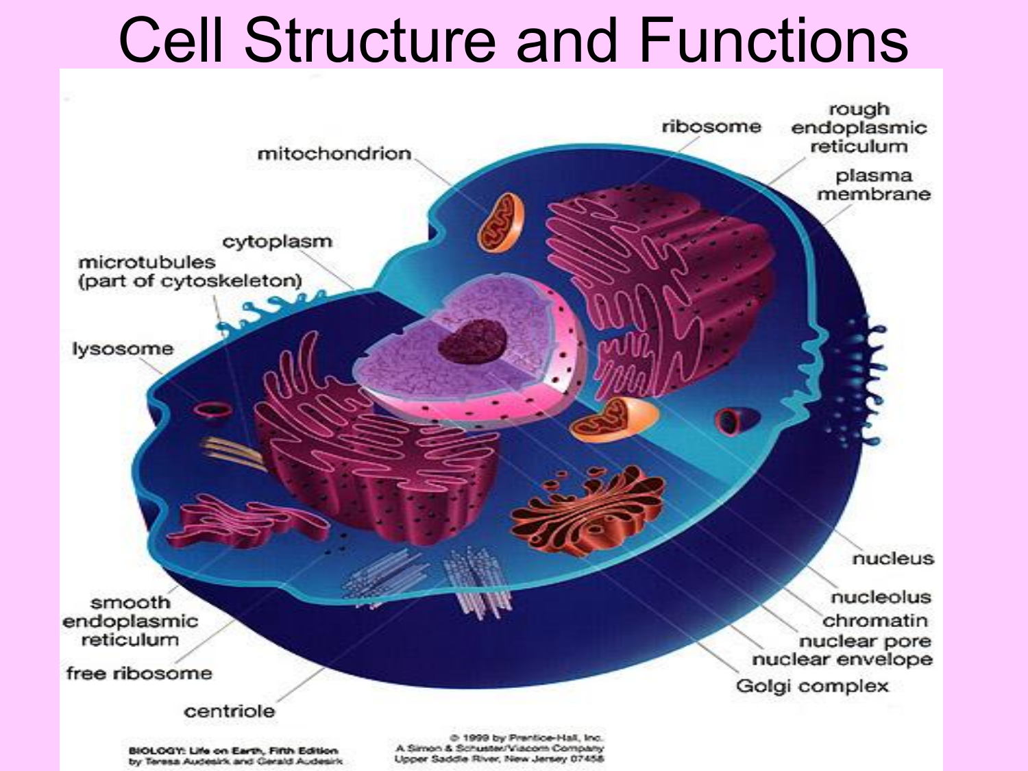
Cell Structure and Functions
The interior of human cells is divided into the nucleus and the cytoplasm.The nucleus is a spherical or oval-shaped structure at the center of the cell. The cytoplasm is the region outside the nucleus that contains cell organelles and cytosol, or cytoplasmic solution.Intracellular fluid is collectively the cytosol and the fluid inside the organelles and the cell nucleus.

The Human Cell Atlas An international effort
The diagram shows five levels of organization in a multicellular organism. The most basic unit is the cell; groups of similar cells form tissues; groups of different tissues make up organs; groups of organs form organ systems; cells, tissues, organs, and organ systems combine to form a multicellular organism.. The human body consists of.
Eukaryotic Plant Cell Organelles / Plant Cell Structures ( Read
ADVERTISEMENTS: Let us make an in-depth study of the structure and functions of cell. After reading this article you will learn about: 1. Comparison of Prokaryotic Cells and Eukaryotic Cells and 2. Structure and Components of a Human Cell. Cell is a compartment where all the activities of life takes place. There are two basic […]

Cell Structure And Function Cells The Basic Units Of Life Siyavula
An Introduction to the Human Body. 1.0 Introduction. 1.1 How Structure Determines Function. 1.2 Structural Organization of the Human Body. 1.3 Homeostasis. 1.4 Anatomical Terminology.. The cell membrane is an extremely pliable structure composed primarily of two layers of phospholipids (a "bilayer"). Cholesterol and various proteins are.